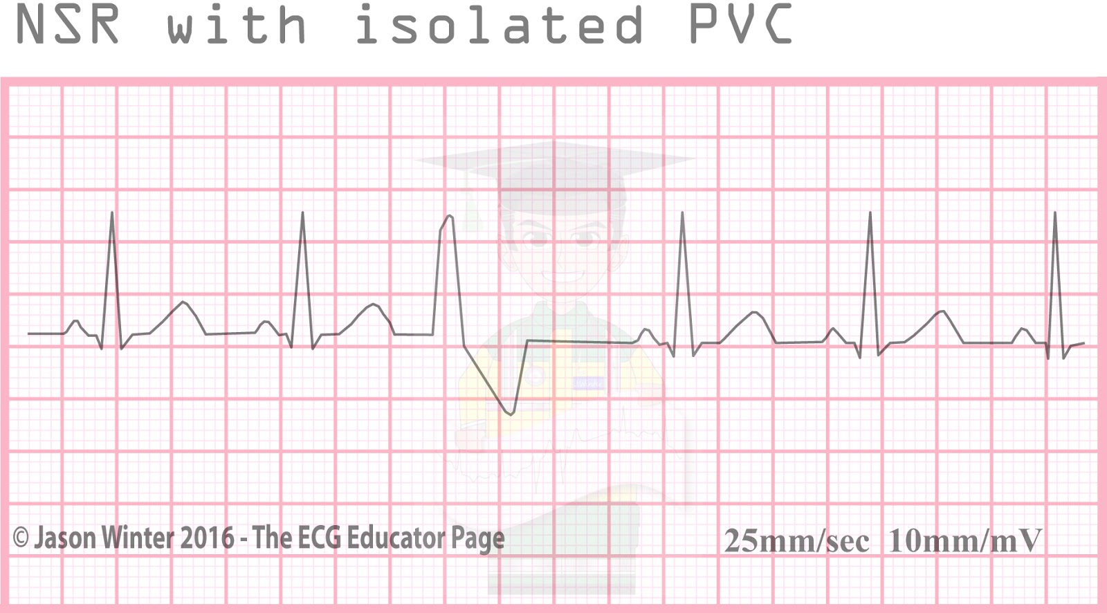
With full compensatory pause, next normal beat arrives an interval is equal double preceding R-R interval. Retrograde capture describes process the ectopic impulse conducted retrogradely the AV node, producing atrial depolarisation. is visible the ECG an inverted P wave ("retrograde P wave"), occurring the QRS complex.
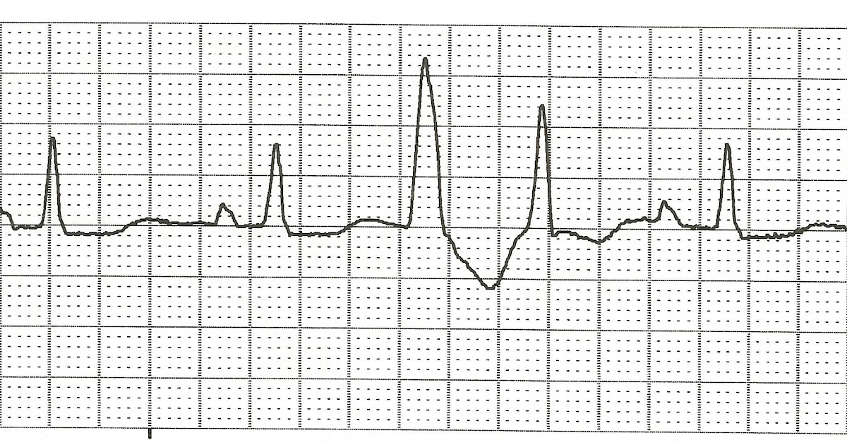 A typical PVC appears an ECG a wide bizarre QRS complex, occurring within underlying normal sinus rhythm. instance, is example an underlying rhythm a normal sinus rhythm a premature ventricular contraction the fourth beat followed this beat a compensatory pause. pause the .
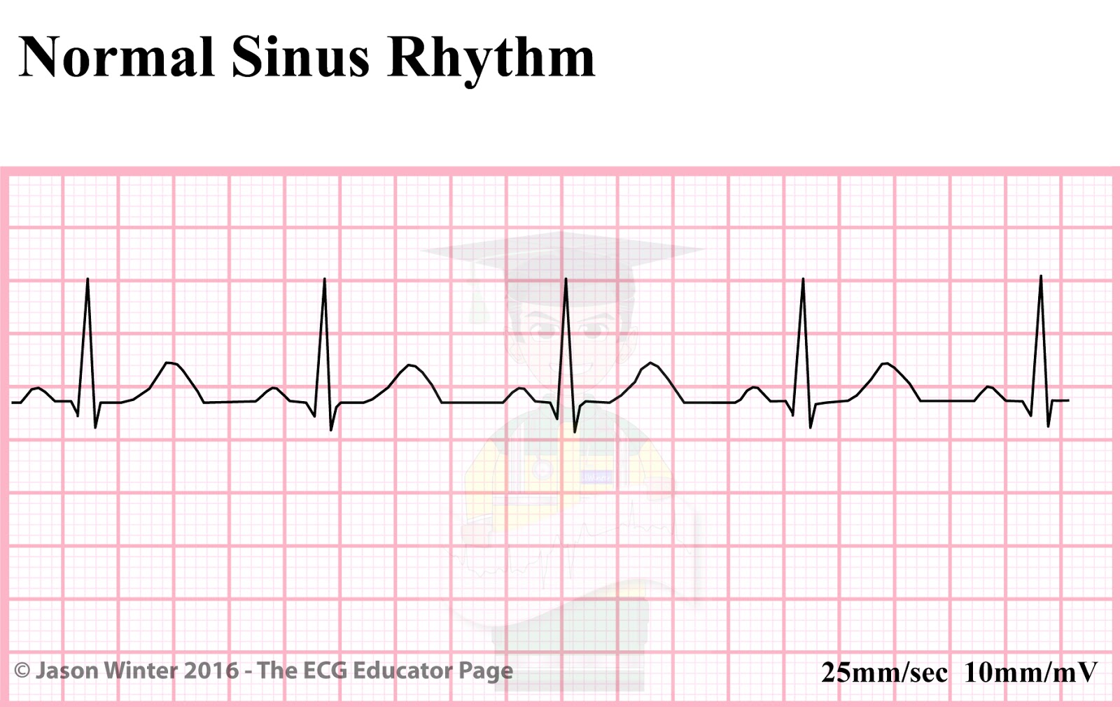
 This ECG shows underlying rhythm normal sinus rhythm a rate 80 / min. are premature ventricular contractions (PVCs). sinus rhythm continues uninterrupted, causing "compensatory pause". you march the P waves, may see hints the hidden P waves the ST segments the PVCs.
 Two consecutive premature ventricular contractions referred as pair couplet.If 3 30 premature ventricular contractions occur consecutively, is referred as non-sustained ventricular tachycardia (if rate >100 beats/min) ventricular rhythm (if rate <100 beats/min). more 30 consecutive beats premature ventricular contractions is referred as .
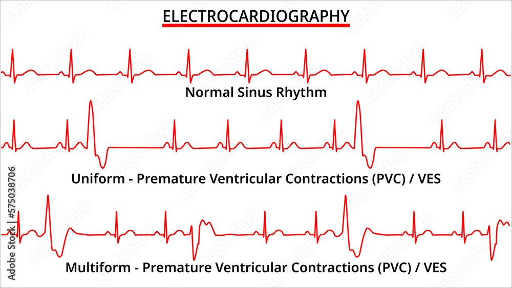
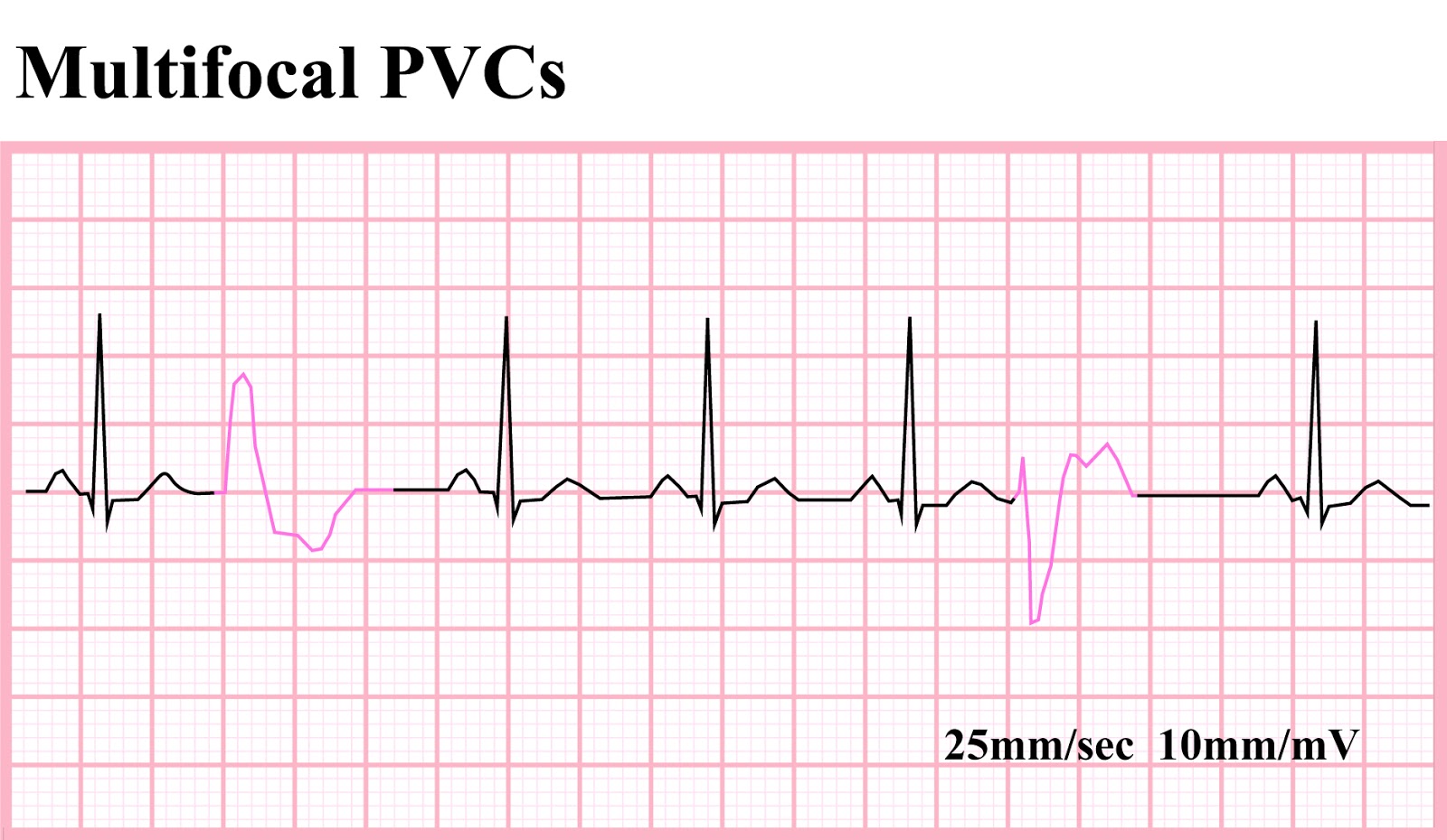
 The morphology the PVC vary the ECG Rhythm more one area the ventricles irritable spontaneously discharge, designated multifocal mulitiform PVCs. suggests more irritable ventricle sometimes lead the rhythm Ventricular Tachycardia. occurrence may trigger .

 ECG Educator Blog : Sinoatrial Node rhythms
 Note: strip appears be NSR on 7th beat, the right, have PVC. Notice the QRS complex the PVC wide stretched out. ectopic nature PVCs only early contraction the ventricles also causes ventricular beats be abnormal. So, pictured is basic singular PVC.
 This ECG shows underlying rhythm normal sinus rhythm a rate 80 / min. are premature ventricular contractions (PVCs). sinus rhythm continues uninterrupted, causing "compensatory pause". you march the P waves, may see hints the hidden P waves the ST segments the PVCs.
 Rhythm analysis normal sinus rhythm (NSR) 84 bpm. Frequent premature ventricular contractions (PVCs) present. encounter shows normal sinus rhythm frequent premature ventricular contractions. PVCs identified a wide, bizarre QRS complex. Frequent PVCs develop arrhythmias cardiomyopathy.
 PVC ECG Strip. Analyze tracing the steps rhythm analysis. Show Answer. Rhythm: Irregular; Rate: 60; P Wave: upright uniform, absent early complex; PR interval: 0.16 sec; QRS: 0.08 sec, early complex wide bizarre 0.16 sec; Interpretation: Sinus Rhythm (NSR) with PVC
 Premature Ventricular Contractions (PVCs) ECG (Example 2) | Learn the Heart

 Premature Ventricular Contractions Pvcs Example 1 | Images and Photos
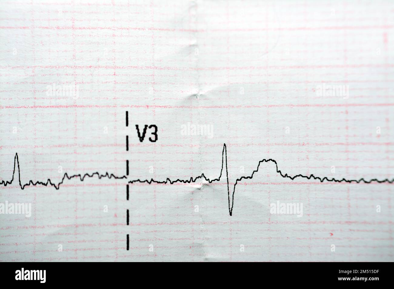
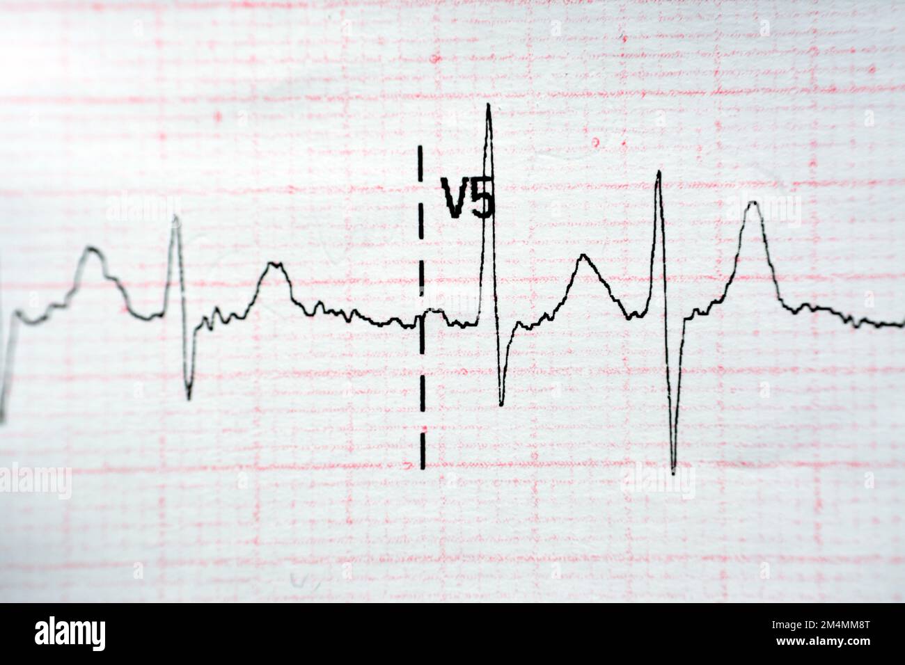 ECG ElectroCardioGraph paper that shows Normal Sinus Rhythm NSR with
 ECG Educator Blog : Ventricular Ectopics
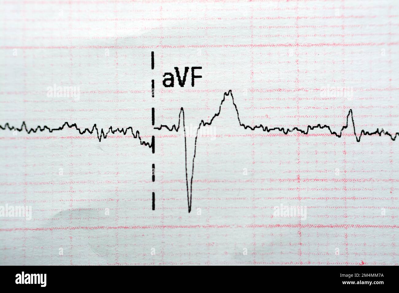 ECG ElectroCardioGraph paper that shows Normal Sinus Rhythm NSR with
 ECG ElectroCardioGraph Paper that Shows Normal Sinus Rhythm NSR with
 pvc-sinus-rhythm - Cardiac Sciences Manitoba
 EKG Rhythm Strips 72
 Practice EKG Rhythm Strips 137

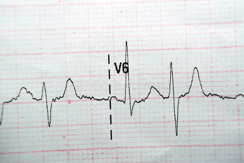 ECG ElectroCardioGraph Paper that Shows Normal Sinus Rhythm NSR with
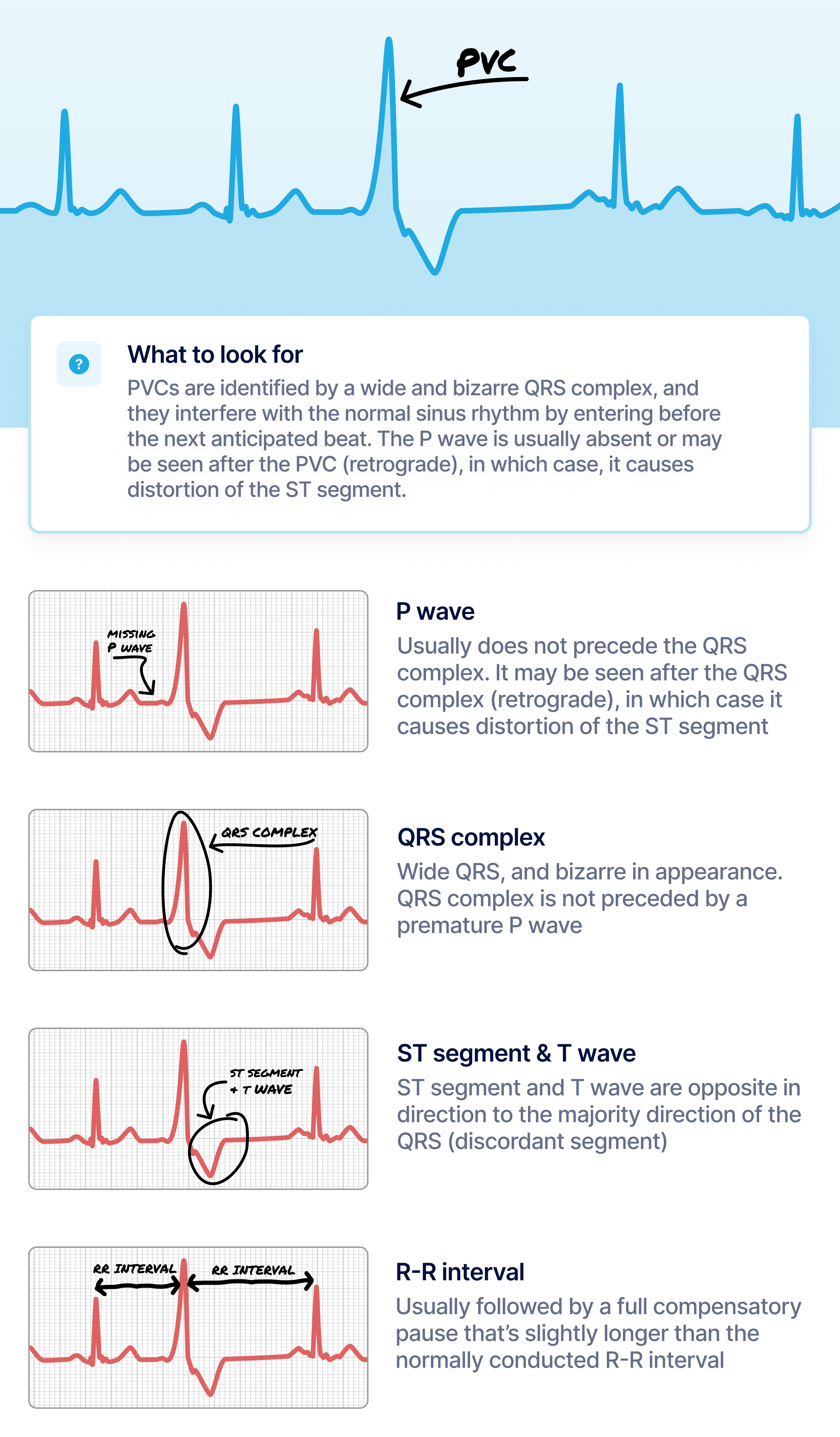 What Premature Ventricular Contraction (PVC) Looks Like on Your Watch
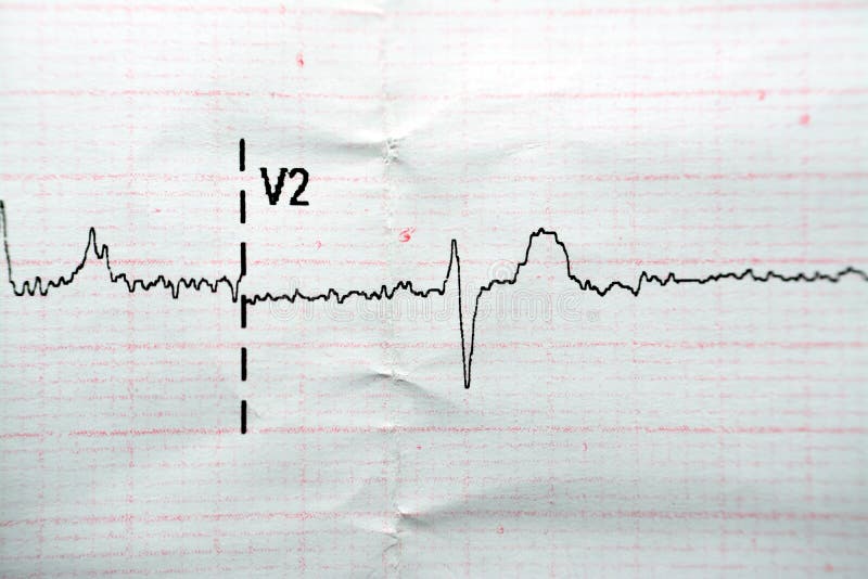 ECG ElectroCardioGraph Paper that Shows Normal Sinus Rhythm NSR with
 ECG ElectroCardioGraph Paper that Shows Normal Sinus Rhythm NSR with
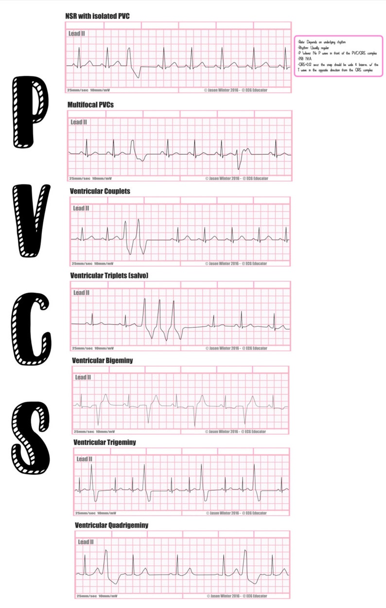 EKG Need to Knows/ Nursing/ EMT Paramedic/ Cardiology - Etsy
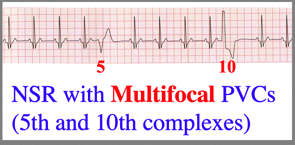 More PVC configurations
 What Premature Ventricular Contraction (PVC) Looks Like on Your Watch
 Advanced EKGs - PACs and PVCs (ie premature beats) - YouTube
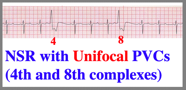 More PVC configurations
 Pvc Ekg Types
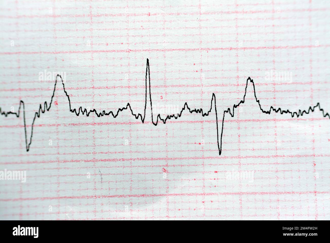 ECG ElectroCardioGraph paper that shows Normal Sinus Rhythm NSR with
 Premature Ventricular Contractions (PVCs) ECG Review | Learn the Heart
 Examples of ECG signal with the most common QRS types: normal sinus
